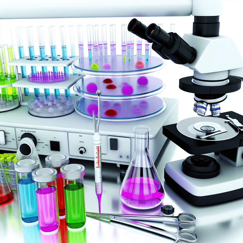
Discover the latest cutting-edge tools revolutionizing cell biology research and unlocking new insights into the building blocks of life.
Cutting-Edge Tools Transforming Cell Biology Research

Table of Contents
- Introduction
- Advancements in Single-Cell Analysis Techniques for Studying Cellular Heterogeneity
- The Role of CRISPR-Cas9 in Revolutionizing Gene Editing and Manipulation in Cell Biology
- Emerging Microscopy Technologies for Visualizing Subcellular Structures and Processes
- The Impact of Artificial Intelligence and Machine Learning on Cell Biology Research and Data Analysis
- Q&A
- Conclusion
“Revolutionize your cell biology research with cutting-edge tools.”
Introduction
Cell biology research has been revolutionized by the development of cutting-edge tools that allow scientists to study cells in unprecedented detail. These tools have enabled researchers to delve deeper into the inner workings of cells, uncovering new insights and advancing our understanding of complex biological processes. From advanced imaging techniques to powerful genetic engineering tools, these cutting-edge technologies are transforming the field of cell biology and paving the way for groundbreaking discoveries. In this article, we will explore some of the most exciting and innovative tools that are driving the progress of cell biology research.
Advancements in Single-Cell Analysis Techniques for Studying Cellular Heterogeneity
Cell biology research has come a long way since the discovery of the cell in the 17th century. With advancements in technology and techniques, scientists are now able to study cells at a level of detail that was once unimaginable. One of the most exciting areas of research in cell biology is the study of cellular heterogeneity, which refers to the differences between individual cells within a population. This heterogeneity plays a crucial role in various biological processes, such as development, disease progression, and response to treatment. In this article, we will explore the cutting-edge tools that are transforming cell biology research, specifically in the field of single-cell analysis techniques for studying cellular heterogeneity.
Traditionally, cell biology research has relied on bulk analysis, where a large number of cells are analyzed together, masking any differences between individual cells. However, with the advent of single-cell analysis techniques, scientists can now study the heterogeneity within a population of cells. This has opened up a whole new world of possibilities for understanding the complex nature of cells and their functions.
One of the most significant advancements in single-cell analysis techniques is the development of single-cell RNA sequencing (scRNA-seq). This technique allows researchers to analyze the gene expression of individual cells, providing insights into the molecular mechanisms that govern cellular heterogeneity. With scRNA-seq, scientists can identify rare cell types, characterize cell subpopulations, and track changes in gene expression over time. This has been particularly useful in cancer research, where tumor heterogeneity is a major challenge in developing effective treatments.
Another cutting-edge tool in single-cell analysis is mass cytometry, also known as CyTOF (Cytometry by Time-Of-Flight). This technique combines the high-throughput capabilities of flow cytometry with the high-resolution capabilities of mass spectrometry. Mass cytometry allows for the simultaneous measurement of multiple parameters in a single cell, providing a more comprehensive view of cellular heterogeneity. This has been instrumental in understanding the immune system, where different cell types and their functions can be identified and characterized.
In addition to these techniques, advancements in imaging technologies have also revolutionized single-cell analysis. Super-resolution microscopy, for example, allows for the visualization of cellular structures and processes at a resolution that was previously unattainable. This has been crucial in studying the spatial organization of cells and how it relates to cellular heterogeneity. Furthermore, live-cell imaging techniques, such as time-lapse microscopy, have enabled researchers to track the behavior of individual cells over time, providing insights into dynamic processes such as cell division and migration.
The integration of these cutting-edge tools has also led to the development of multi-omics approaches, where multiple types of data, such as genomic, transcriptomic, and proteomic, can be analyzed from the same single cell. This has been a game-changer in understanding the complex interactions between different cellular components and how they contribute to cellular heterogeneity. Multi-omics approaches have been particularly useful in studying stem cells, where the ability to differentiate into different cell types is governed by complex molecular networks.
The advancements in single-cell analysis techniques have not only transformed basic research but also have significant implications in clinical applications. For instance, single-cell analysis has been instrumental in the development of personalized medicine, where treatments can be tailored to an individual’s unique cellular makeup. This has been particularly beneficial in cancer treatment, where the heterogeneity of tumors makes it challenging to develop effective therapies.
In conclusion, the cutting-edge tools discussed in this article have revolutionized the study of cellular heterogeneity and have opened up new avenues for understanding the complex nature of cells. With the integration of these techniques, scientists can now study cells at a level of detail that was once unimaginable, providing insights into various biological processes and their implications in health and disease. As technology continues to advance, we can only expect further breakthroughs in single-cell analysis techniques, leading to a deeper understanding of the fundamental building blocks of life – the cells.
The Role of CRISPR-Cas9 in Revolutionizing Gene Editing and Manipulation in Cell Biology

Cell biology research has come a long way in recent years, thanks to the development of cutting-edge tools and technologies. One such tool that has revolutionized the field is CRISPR-Cas9, a gene editing and manipulation tool that has opened up new possibilities for researchers.
CRISPR-Cas9, short for Clustered Regularly Interspaced Short Palindromic Repeats-CRISPR associated protein 9, is a bacterial defense system that has been adapted for use in gene editing. It was first discovered in 1987 by Japanese researchers, but it wasn’t until 2012 that its potential for gene editing was realized by a team of scientists led by Jennifer Doudna and Emmanuelle Charpentier.
The CRISPR-Cas9 system works by using a guide RNA to target a specific sequence of DNA, and then using the Cas9 enzyme to cut the DNA at that location. This allows for precise and efficient editing of the genetic code, making it a valuable tool for cell biology research.
One of the key advantages of CRISPR-Cas9 is its simplicity and ease of use. Traditional gene editing techniques, such as zinc finger nucleases and TALENs, require a lot of time and expertise to design and implement. CRISPR-Cas9, on the other hand, can be easily programmed to target any desired sequence of DNA, making it accessible to a wider range of researchers.
In addition to its ease of use, CRISPR-Cas9 also offers a high level of precision. This is due to the fact that the guide RNA can be designed to match a specific sequence of DNA, minimizing the risk of off-target effects. This precision is crucial in cell biology research, where even small changes in the genetic code can have significant impacts on cellular function.
The potential applications of CRISPR-Cas9 in cell biology research are vast. One of the most exciting areas of research is in the study of disease. By using CRISPR-Cas9 to edit the genetic code of cells, researchers can create disease models that closely mimic human conditions. This allows for a better understanding of the underlying mechanisms of diseases and the development of potential treatments.
Another area where CRISPR-Cas9 is making a significant impact is in the field of regenerative medicine. By using the tool to edit the genetic code of stem cells, researchers can create customized cells that can be used for tissue repair and regeneration. This has the potential to revolutionize the treatment of diseases and injuries that currently have limited treatment options.
CRISPR-Cas9 is also being used to study the function of specific genes in cell biology. By knocking out or modifying a gene of interest, researchers can observe the effects on cellular processes and gain a better understanding of their function. This has already led to significant discoveries in areas such as cancer research and developmental biology.
However, as with any new technology, there are also ethical concerns surrounding the use of CRISPR-Cas9. The potential for unintended consequences and the ability to manipulate the genetic code of living organisms raises questions about the ethical implications of its use. As such, it is crucial for researchers to use this tool responsibly and ethically.
In conclusion, CRISPR-Cas9 has emerged as a game-changing tool in cell biology research. Its simplicity, precision, and versatility have opened up new possibilities for studying diseases, developing treatments, and understanding the fundamental processes of life. As research in this field continues to advance, it is clear that CRISPR-Cas9 will play a crucial role in shaping the future of cell biology.
Emerging Microscopy Technologies for Visualizing Subcellular Structures and Processes
Cell biology research has come a long way since the invention of the microscope in the 17th century. With the advancement of technology, scientists now have access to cutting-edge tools that allow them to visualize subcellular structures and processes in unprecedented detail. These emerging microscopy technologies have revolutionized the field of cell biology, providing researchers with a deeper understanding of cellular functions and paving the way for new discoveries.
One of the most exciting developments in microscopy is the use of super-resolution techniques. Traditional light microscopes are limited by the diffraction of light, which prevents the visualization of structures smaller than 200 nanometers. Super-resolution microscopy overcomes this limitation by using specialized techniques to bypass the diffraction barrier. This allows for the visualization of structures as small as 20 nanometers, providing researchers with a level of detail that was previously unattainable.
One type of super-resolution microscopy is stimulated emission depletion (STED) microscopy. This technique uses two laser beams, one to excite the fluorescent molecules and another to deplete the fluorescence in a specific area. By scanning the sample with the two beams, a high-resolution image can be reconstructed. STED microscopy has been used to study the organization of proteins in the cell membrane and to visualize the dynamics of cellular processes such as endocytosis.
Another super-resolution technique is structured illumination microscopy (SIM). This method uses patterned light to illuminate the sample, which is then reconstructed into a high-resolution image. SIM has been used to study the structure and function of cilia, the hair-like structures on the surface of cells that play a crucial role in cell movement and signaling.
In addition to super-resolution techniques, advances in electron microscopy have also transformed cell biology research. Transmission electron microscopy (TEM) has been used for decades to visualize subcellular structures at high resolution. However, recent developments in cryo-electron microscopy (cryo-EM) have allowed for the visualization of biological samples in their native state, without the need for chemical fixation or staining. This has provided researchers with a more accurate representation of cellular structures and processes.
Cryo-EM has also been combined with tomography, a technique that allows for the reconstruction of three-dimensional images from a series of two-dimensional images. This has enabled researchers to study the 3D organization of cellular structures and to visualize dynamic processes such as cell division and protein transport.
In addition to these advanced microscopy techniques, new imaging probes and labels have also been developed to enhance the visualization of subcellular structures. Fluorescent proteins, such as green fluorescent protein (GFP), have been widely used to label specific proteins and track their movements within the cell. However, these proteins have limitations in terms of brightness and photostability. To overcome these limitations, new fluorescent probes have been developed, such as quantum dots and organic dyes, which provide brighter and more stable signals.
Furthermore, the development of genetically encoded sensors has allowed for the visualization of specific cellular processes, such as calcium signaling and protein-protein interactions. These sensors can be targeted to specific organelles or cellular structures, providing researchers with a more precise understanding of their functions.
In conclusion, the emergence of these cutting-edge tools has transformed the field of cell biology, allowing researchers to visualize subcellular structures and processes in unprecedented detail. Super-resolution techniques, advances in electron microscopy, and the development of new imaging probes and sensors have provided researchers with a deeper understanding of cellular functions and have opened up new avenues for research. As technology continues to advance, we can expect even more exciting developments in the field of cell biology, leading to new discoveries and breakthroughs in our understanding of life at the cellular level.
The Impact of Artificial Intelligence and Machine Learning on Cell Biology Research and Data Analysis
Cell biology research has come a long way since the discovery of the cell in the 17th century. With advancements in technology and the development of cutting-edge tools, scientists are now able to delve deeper into the intricate world of cells and their functions. One of the most significant developments in recent years has been the integration of artificial intelligence (AI) and machine learning (ML) in cell biology research and data analysis.
AI and ML are revolutionizing the way scientists study cells by providing powerful tools for data analysis and interpretation. These technologies have the ability to analyze vast amounts of data in a fraction of the time it would take a human researcher, making it possible to uncover patterns and relationships that would have been impossible to detect otherwise.
One of the key areas where AI and ML are making a significant impact is in image analysis. Microscopy has long been a crucial tool in cell biology research, allowing scientists to visualize and study cells at a microscopic level. However, analyzing the vast amount of data generated by microscopy can be a time-consuming and labor-intensive process. With the help of AI and ML, this process has been streamlined, allowing for faster and more accurate analysis of images.
AI and ML algorithms can be trained to recognize and classify different cell types, structures, and patterns in images. This not only saves time but also reduces the potential for human error. Additionally, these algorithms can learn and improve over time, making them even more efficient and accurate.
Another area where AI and ML are transforming cell biology research is in data analysis. With the advent of high-throughput technologies, such as next-generation sequencing, researchers are now able to generate vast amounts of data in a short period. However, analyzing this data and extracting meaningful insights can be a daunting task.
AI and ML algorithms can handle large datasets with ease, making it possible to identify patterns and relationships that would have been missed by traditional methods. These algorithms can also be used to predict outcomes and make connections between different datasets, providing valuable insights into the complex world of cells.
One of the most exciting applications of AI and ML in cell biology research is in drug discovery. Developing new drugs is a lengthy and expensive process, with a high failure rate. However, with the help of AI and ML, researchers can now screen thousands of compounds and predict their potential efficacy and toxicity, saving time and resources.
AI and ML can also be used to identify new drug targets by analyzing large datasets and identifying patterns that may not be apparent to human researchers. This has the potential to revolutionize the drug discovery process and lead to the development of more effective treatments for various diseases.
In addition to aiding in data analysis, AI and ML are also being used to design experiments and optimize protocols. These technologies can analyze past experiments and suggest improvements, leading to more efficient and effective research.
However, as with any new technology, there are also challenges and limitations to consider. One of the main concerns with the use of AI and ML in cell biology research is the potential for bias. These algorithms are only as good as the data they are trained on, and if the data is biased, the results will be as well. Therefore, it is crucial for researchers to carefully select and validate their data to ensure the accuracy and reliability of their findings.
In conclusion, the integration of AI and ML in cell biology research and data analysis is transforming the field in unprecedented ways. These technologies are providing powerful tools for image analysis, data analysis, and drug discovery, making it possible to uncover new insights and advance our understanding of cells. While there are challenges to consider, the potential for these technologies to revolutionize cell biology research is undeniable. As we continue to push the boundaries of what is possible, the future of cell biology research looks brighter than ever before.
Q&A
1. What are some examples of cutting-edge tools used in cell biology research?
Some examples of cutting-edge tools used in cell biology research include CRISPR-Cas9 gene editing technology, single-cell sequencing techniques, super-resolution microscopy, and optogenetics.
2. How has CRISPR-Cas9 technology transformed cell biology research?
CRISPR-Cas9 technology has revolutionized cell biology research by allowing scientists to easily and precisely edit the genetic code of cells. This has opened up new possibilities for studying the function of specific genes and their role in diseases.
3. What is single-cell sequencing and how is it being used in cell biology research?
Single-cell sequencing is a technique that allows researchers to analyze the genetic material of individual cells, rather than a mixture of cells. This has enabled a deeper understanding of cellular heterogeneity and has been used to study complex diseases such as cancer.
4. What is optogenetics and how is it being used in cell biology research?
Optogenetics is a technique that uses light to control the activity of specific cells in living organisms. It has been used in cell biology research to study the function of neurons and other cells, and has potential applications in treating neurological disorders.
Conclusion
In conclusion, cutting-edge tools have greatly transformed cell biology research by providing scientists with advanced techniques and technologies to study cells at a molecular level. These tools have allowed for a deeper understanding of cellular processes and have led to groundbreaking discoveries in the field. With the continuous development of new and innovative tools, the future of cell biology research looks promising and will continue to push the boundaries of our knowledge about the fundamental unit of life. These advancements have the potential to revolutionize the way we approach and treat diseases, ultimately improving human health and well-being. Overall, cutting-edge tools are essential for the progress of cell biology research and will continue to play a crucial role in shaping the future of this field.


