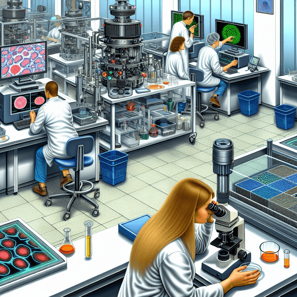
Discover the latest advancements in cellular imaging and analysis with emerging technologies. Stay ahead of the curve in this rapidly evolving field.
Emerging Technologies in Cellular Imaging and Analysis

Table of Contents
“Unleashing the power of technology to revolutionize cellular imaging and analysis.”
Introduction
Emerging technologies in cellular imaging and analysis have revolutionized the field of biology and medicine. These cutting-edge techniques allow scientists and researchers to visualize and analyze cells at a level of detail that was previously unimaginable. By combining advanced imaging technologies with powerful analytical tools, researchers are able to gain a deeper understanding of cellular structures and functions, leading to new discoveries and advancements in various fields such as cancer research, drug development, and regenerative medicine. In this introduction, we will explore some of the most exciting emerging technologies in cellular imaging and analysis and their potential impact on the future of scientific research.
Advancements in Fluorescence Microscopy for Cellular Imaging
Cellular imaging and analysis have revolutionized the field of biology and medicine, allowing researchers and clinicians to visualize and study cells in unprecedented detail. Over the years, there have been significant advancements in imaging technologies, with fluorescence microscopy being one of the most widely used techniques. This article will delve into the emerging technologies in fluorescence microscopy for cellular imaging and analysis.
Fluorescence microscopy is a powerful tool that utilizes fluorescent molecules to label specific structures or molecules within cells. These molecules emit light of a specific wavelength when excited by a light source, allowing for the visualization of cellular structures and processes. Traditional fluorescence microscopy techniques, such as widefield and confocal microscopy, have been instrumental in advancing our understanding of cellular biology. However, with the rapid pace of technological advancements, new and improved techniques have emerged, providing researchers with even more detailed and accurate imaging capabilities.
One of the most significant advancements in fluorescence microscopy is the development of super-resolution microscopy. This technique overcomes the diffraction limit of light, which was previously thought to be the ultimate resolution limit in microscopy. Super-resolution microscopy techniques, such as stimulated emission depletion (STED) microscopy and structured illumination microscopy (SIM), use specialized optics and algorithms to achieve resolutions up to 10 times higher than traditional fluorescence microscopy. This has allowed researchers to visualize cellular structures and processes at the nanoscale, providing insights into previously unobservable details.
Another emerging technology in fluorescence microscopy is single-molecule localization microscopy (SMLM). This technique utilizes the stochastic switching of fluorescent molecules to achieve super-resolution imaging. By precisely localizing individual molecules, SMLM can achieve resolutions down to a few nanometers. This has been particularly useful in studying the organization and dynamics of cellular structures, such as the cytoskeleton and membrane proteins. SMLM has also been combined with other techniques, such as fluorescence resonance energy transfer (FRET), to study protein-protein interactions within cells.
In addition to advancements in resolution, there have also been significant improvements in imaging speed and sensitivity. High-speed fluorescence microscopy techniques, such as light-sheet microscopy and spinning disk confocal microscopy, have enabled the visualization of dynamic cellular processes in real-time. These techniques use specialized illumination and detection methods to capture images at high speeds, allowing for the study of fast-moving cellular structures and processes. Furthermore, the development of highly sensitive cameras and detectors has improved the signal-to-noise ratio in fluorescence microscopy, enabling the detection of even faint fluorescent signals.
The integration of artificial intelligence (AI) and machine learning (ML) in fluorescence microscopy has also been a game-changer in cellular imaging and analysis. These technologies have been used to automate and streamline image analysis, reducing the time and effort required for data analysis. AI and ML algorithms can identify and quantify cellular structures and processes, such as cell morphology and protein localization, with high accuracy and efficiency. This has not only improved the speed and accuracy of data analysis but has also allowed for the analysis of large datasets, providing a more comprehensive understanding of cellular biology.
In conclusion, the advancements in fluorescence microscopy have greatly enhanced our ability to visualize and study cells. From super-resolution imaging to high-speed and sensitive techniques, these emerging technologies have provided researchers with unprecedented capabilities in cellular imaging and analysis. The integration of AI and ML has further improved the efficiency and accuracy of data analysis, paving the way for new discoveries in the field of biology and medicine. As technology continues to advance, we can only imagine the possibilities and potential breakthroughs that lie ahead in cellular imaging and analysis.
The Role of Artificial Intelligence in Automated Cell Analysis

Cellular imaging and analysis have been crucial tools in the field of biology and medicine for decades. With the advancement of technology, these tools have become more sophisticated and accurate, allowing researchers to gain a deeper understanding of cellular processes and diseases. One of the most exciting developments in this field is the integration of artificial intelligence (AI) in automated cell analysis.
AI is a branch of computer science that focuses on creating intelligent machines that can perform tasks that typically require human intelligence. In the context of cellular imaging and analysis, AI algorithms are used to analyze large amounts of data and identify patterns and abnormalities that may not be easily detectable by the human eye. This has revolutionized the way researchers study cells and has opened up new possibilities for diagnosis and treatment of diseases.
One of the main advantages of using AI in automated cell analysis is its ability to process vast amounts of data in a short period. Traditional methods of cell analysis involve manually counting and analyzing cells, which is a time-consuming and tedious process. With AI, this task can be completed in a fraction of the time, allowing researchers to focus on other aspects of their research.
Moreover, AI algorithms can analyze data with a level of accuracy and consistency that is not achievable by humans. This is especially beneficial in cases where the cells being analyzed are small or have subtle differences. AI can detect these differences and patterns that may not be visible to the human eye, providing researchers with a more comprehensive understanding of cellular processes.
Another significant advantage of using AI in automated cell analysis is its ability to learn and adapt. As the algorithm is exposed to more data, it can continuously improve its accuracy and efficiency. This means that the more data it analyzes, the better it becomes at identifying patterns and abnormalities. This is particularly useful in the field of medicine, where diseases can present themselves in various ways, and having a tool that can adapt to these changes is invaluable.
One area where AI has shown great potential is in the diagnosis of diseases. With the help of AI, researchers can analyze images of cells and tissues to identify abnormalities that may indicate the presence of a disease. This has the potential to revolutionize the way diseases are diagnosed, as it can provide more accurate and timely results. Moreover, AI can also assist in predicting the progression of diseases, allowing for early intervention and treatment.
In addition to diagnosis, AI can also aid in drug discovery and development. By analyzing the effects of different compounds on cells, AI algorithms can identify potential drug candidates and predict their efficacy. This can significantly speed up the drug development process, which is often a lengthy and costly endeavor.
However, like any technology, AI also has its limitations. One of the main challenges is the lack of diversity in the data used to train the algorithms. This can lead to biased results and hinder the algorithm’s ability to accurately analyze data from different populations. To overcome this, researchers must ensure that the data used to train AI algorithms is diverse and representative of the population being studied.
In conclusion, the integration of AI in automated cell analysis has opened up new possibilities in the field of cellular imaging and analysis. Its ability to process vast amounts of data, identify patterns and abnormalities, and continuously learn and adapt has made it an invaluable tool for researchers. As technology continues to advance, we can expect to see even more significant developments in this field, leading to a deeper understanding of cellular processes and improved diagnosis and treatment of diseases.
Exploring the Potential of Single-Cell Analysis in Disease Research
Cellular imaging and analysis have been crucial tools in disease research for decades. However, with the rapid advancements in technology, there has been a shift towards single-cell analysis, which allows for a more detailed and precise understanding of diseases at the cellular level. This emerging technology has the potential to revolutionize disease research and pave the way for more effective treatments.
Traditionally, cellular imaging and analysis involved studying a large population of cells and averaging out the results. This approach has its limitations as it does not take into account the heterogeneity of cells within a population. With single-cell analysis, researchers can now study individual cells and gain a deeper understanding of their behavior and function.
One of the most significant advantages of single-cell analysis is the ability to identify rare cell types. In diseases such as cancer, there may be a small population of cells that are responsible for driving the progression of the disease. These cells may be missed when studying a large population, but with single-cell analysis, they can be identified and studied in detail. This can lead to the development of targeted therapies that specifically target these rare cells, improving treatment outcomes.
Another area where single-cell analysis is making a significant impact is in the study of the immune system. The immune system is a complex network of cells that work together to protect the body from foreign invaders. With single-cell analysis, researchers can now study the different types of immune cells and their interactions in response to diseases. This has led to a better understanding of immune dysfunction in diseases such as autoimmune disorders and cancer.
In addition to identifying rare cell types, single-cell analysis also allows for the study of cellular heterogeneity. Within a population of cells, there can be significant differences in gene expression, protein levels, and other cellular characteristics. These differences can have a significant impact on disease progression and treatment response. With single-cell analysis, researchers can now study these differences and gain a more comprehensive understanding of disease mechanisms.
One of the most exciting applications of single-cell analysis is in the field of personalized medicine. With this technology, researchers can study individual cells from a patient and tailor treatment plans based on their specific cellular characteristics. This has the potential to improve treatment outcomes and reduce the risk of adverse reactions to medications.
The advancements in single-cell analysis have also led to the development of new imaging techniques. Traditional imaging methods, such as fluorescence microscopy, have limitations in terms of resolution and sensitivity. However, with the development of super-resolution microscopy and single-molecule imaging, researchers can now visualize cellular structures and processes at a much higher resolution. This has opened up new possibilities for studying diseases at the molecular level.
Despite the numerous advantages of single-cell analysis, there are still challenges that need to be addressed. One of the main challenges is the handling and processing of individual cells. As the name suggests, single-cell analysis involves studying individual cells, which can be time-consuming and labor-intensive. There is also a need for standardized protocols and data analysis methods to ensure consistency and reproducibility of results.
In conclusion, single-cell analysis is an emerging technology that has the potential to transform disease research. It allows for the identification of rare cell types, study of cellular heterogeneity, and development of personalized treatment plans. With the continued advancements in technology, we can expect to see even more exciting developments in this field, leading to a better understanding of diseases and improved treatment options.
Innovations in Live Cell Imaging Techniques for Real-Time Analysis
Cellular imaging and analysis have revolutionized the field of biology and medicine, allowing researchers to observe and understand the intricate processes that occur within living cells. With the advancement of technology, new and innovative techniques have emerged, providing scientists with the ability to capture real-time images and analyze cellular behavior in unprecedented detail. In this article, we will explore some of the emerging technologies in cellular imaging and analysis, specifically focusing on the latest innovations in live cell imaging techniques.
One of the most significant developments in live cell imaging is the use of fluorescent probes and dyes. These molecules can be specifically targeted to different cellular structures or molecules, allowing researchers to visualize and track their movements in real-time. This technique has been particularly useful in studying dynamic processes such as cell division, protein trafficking, and signaling pathways.
Another emerging technology in live cell imaging is the use of genetically encoded fluorescent proteins. These proteins can be genetically engineered to fuse with specific cellular structures or proteins, providing a more precise and long-term visualization of their behavior. This technique has been instrumental in studying the dynamics of organelles, such as mitochondria and lysosomes, and their interactions with other cellular components.
In recent years, super-resolution microscopy has also emerged as a powerful tool in live cell imaging. This technique overcomes the diffraction limit of traditional light microscopy, allowing for the visualization of structures as small as a few nanometers. Super-resolution microscopy has been particularly useful in studying the organization and dynamics of subcellular structures, such as the cytoskeleton and membrane proteins.
Advancements in imaging hardware have also played a crucial role in the development of live cell imaging techniques. High-speed cameras with improved sensitivity and resolution have enabled the capture of fast-moving cellular processes with high temporal and spatial resolution. Additionally, the development of microfluidic devices has allowed for the precise control of the cellular environment, providing a more physiologically relevant setting for live cell imaging experiments.
One of the most exciting developments in live cell imaging is the integration of imaging techniques with artificial intelligence (AI). AI algorithms can analyze large datasets of live cell images, providing automated and unbiased analysis of cellular behavior. This has significantly reduced the time and effort required for data analysis, allowing researchers to focus on the interpretation of results and the generation of new hypotheses.
Furthermore, the combination of live cell imaging with other techniques, such as single-cell sequencing and mass spectrometry, has opened up new avenues for understanding cellular processes. By integrating multiple techniques, researchers can obtain a more comprehensive and detailed view of cellular behavior, leading to a deeper understanding of complex biological systems.
The emergence of these new technologies has also led to the development of novel applications in live cell imaging. For example, the use of optogenetics, a technique that allows for the control of cellular processes using light, has been integrated with live cell imaging to study the dynamics of cellular signaling pathways. This has provided researchers with a powerful tool to manipulate and study cellular behavior in real-time.
In conclusion, the continuous advancements in technology have revolutionized live cell imaging and analysis, providing researchers with unprecedented capabilities to study cellular processes in real-time. The integration of different techniques and the use of AI has opened up new possibilities for understanding complex biological systems. As these technologies continue to evolve, we can expect to gain even deeper insights into the inner workings of living cells, leading to new discoveries and breakthroughs in the field of biology and medicine.
Q&A
1. What are some examples of emerging technologies in cellular imaging and analysis?
Some examples of emerging technologies in cellular imaging and analysis include super-resolution microscopy, single-cell sequencing, and live-cell imaging using fluorescent probes. Other emerging technologies include machine learning and artificial intelligence for image analysis and 3D imaging techniques such as light sheet microscopy.
2. How do these emerging technologies improve upon traditional methods of cellular imaging and analysis?
These emerging technologies offer higher resolution and sensitivity, allowing for more detailed and accurate imaging of cellular structures and processes. They also enable the study of individual cells and their behavior, providing a deeper understanding of cellular function. Additionally, the use of machine learning and AI can automate and speed up the analysis process, reducing human error and bias.
3. What are some potential applications of these emerging technologies in cellular imaging and analysis?
These emerging technologies have a wide range of potential applications, including drug discovery and development, disease diagnosis and treatment, and basic research in cell biology. They can also be used in fields such as neuroscience, immunology, and cancer research to study the complex interactions and dynamics of cells.
4. Are there any limitations or challenges associated with these emerging technologies?
Some limitations and challenges associated with these emerging technologies include high costs, specialized training and expertise required for operation and analysis, and potential technical issues such as phototoxicity in live-cell imaging. Additionally, the large amount of data generated by these technologies can also pose challenges for storage and analysis.
Conclusion
In conclusion, emerging technologies in cellular imaging and analysis have greatly advanced our understanding of cellular processes and have opened up new possibilities for research and medical applications. From high-resolution imaging techniques to advanced data analysis tools, these technologies have allowed us to visualize and analyze cells in ways that were previously not possible. With continued advancements and integration of these technologies, we can expect to see even more breakthroughs in the field of cellular imaging and analysis, leading to improved diagnostics, treatments, and overall understanding of the complex world of cells. It is an exciting time for this field and the potential for further advancements is endless.


